SolStock/E+/GettyImages
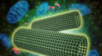
Cell membranes are composed of phospholipid bilayers with embedded proteins that mediate essential cellular functions. Traditional light microscopy cannot resolve individual membrane proteins. Freeze fracture, combined with electron microscopy, allows us to split frozen membranes along their bilayer, exposing protein distribution within the lipid matrix. By integrating additional labeling techniques, researchers can map specific proteins, as well as bacterial and viral components, with sub‑nanometer precision.
1. Rapidly freeze cells or tissues in liquid nitrogen to immobilize structures.
2. Use a microtome to fracture the frozen sample; the membrane cleaves between the two phospholipid leaflets where hydrophobic interactions are weakest.
3. Perform freeze etching in high vacuum to remove ice crystals, preserving fine detail.
4. Shadow the fracture face with a thin film of carbon and platinum to create a stable replica that follows the membrane topology.
5. Digest residual organic material with acid, leaving a platinum shell that represents the exposed membrane surface.
6. Examine the replica by transmission electron microscopy (TEM) for structural analysis.
Freeze etching removes ice crystals that would otherwise obscure membrane detail. The vacuum‑drying process preserves the native architecture and enables observation of dynamic membrane activities, intracellular organelles, and viral assemblies.
Transmission electron microscopy offers up to 1 million‑fold magnification and resolutions down to 3 nm, making it the preferred modality for freeze‑fracture replicas. Scanning electron microscopy (SEM) is less commonly used in this context but can provide surface topology of larger specimens.
Since its introduction over five decades ago, freeze‑fracture TEM has been instrumental in confirming the lipid bilayer model of the plasma membrane and elucidating the spatial arrangement of integral, peripheral, and lipid‑anchored proteins. The technique reveals whether proteins are “floaters” that drift within the bilayer or “anchors” that span both leaflets, and it can detect protein aggregation or clustering. When coupled with immunogold labeling—antibodies tagged with electron‑dense particles—researchers can identify specific proteins and infer their functional roles.