Stocktrek Images/Stocktrek Images/GettyImages
Centrioles are cylindrical, microtubule‑based organelles that sit near the nucleus in most eukaryotic cells. They are essential for accurate chromosome segregation during cell division and for the formation of cilia and flagella. Although absent in prokaryotes, centrioles are a hallmark of animal cells and some lower plants.
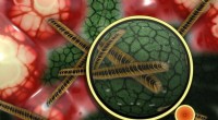
Each centriole consists of nine triplet microtubule bundles radiating from a central cartwheel, forming a right‑angled pair known as the mother–daughter centriole. The cartwheel provides structural rigidity and establishes the nine‑fold symmetry characteristic of centrioles. Surrounding this core is the pericentriolar material (PCM), a protein matrix that nucleates microtubule growth and recruits other centrosomal components.
During interphase, centrioles duplicate to ensure a single pair is available for each daughter cell. In prophase, the centrosomes separate, positioning one centriole pair at each spindle pole. The PCM then organizes the mitotic spindle, a dynamic array of microtubules that capture kinetochores at chromosome centromeres and pull sister chromatids apart during anaphase. This coordinated activity guarantees that each daughter nucleus receives an identical chromosome complement.
Centrioles act as basal bodies, the nucleation sites for axonemal microtubules that compose motile cilia and flagella. The classic “9+2” arrangement—nine outer doublets surrounding two central singlets—arises from centriole‑derived microtubules. Cilia line epithelial surfaces (e.g., the trachea) and move fluids, while flagella, such as the sperm tail, provide motility.
Centrioles are found exclusively in animal cells and in a few lower plant taxa (mosses, liverworts, lichens). Higher plants lack centrioles entirely, instead relying on alternative mechanisms for spindle organization. Even within ciliated cells, the centriole core may be absent, yet the microtubule scaffold remains external.
Mutations in centriole‑associated genes (e.g., OFD1, C2CD3) disrupt ciliary assembly and are linked to ciliopathies such as Oral‑Facial‑Digital (OFD) syndrome and Meckel‑Gruber syndrome. In OFD, patients exhibit craniofacial anomalies, digit malformations, and intellectual disability; the disorder is X‑linked and more prevalent in females. Meckel‑Gruber syndrome presents with renal cysts, brain malformations, and polydactyly, and is autosomal recessive.
Aberrant centriole amplification—extra or enlarged centrioles—is a common hallmark of malignant cells. Loss of the tumor suppressor p53 can bypass centriole‑count checkpoints, leading to chromosomal instability. Targeting centriole over‑duplication has emerged as a potential therapeutic strategy to curb tumor progression.
Ongoing studies aim to decode the regulatory networks that control centriole biogenesis, maturation, and disassembly. A deeper understanding may unlock new avenues for treating ciliopathies and cancers where centriole dysregulation is a driving force.