In both plant and animal cells:
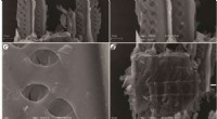
* Nucleus: Contains the genetic material (DNA) of the cell. The electron microscope reveals the nuclear envelope (double membrane), nucleolus (site of ribosome production), and chromatin (DNA and associated proteins).
* Ribosomes: Small, dense particles responsible for protein synthesis. Electron microscopes show their granular structure and their association with the endoplasmic reticulum (ER).
* Endoplasmic Reticulum (ER): An extensive network of interconnected membranes. The electron microscope distinguishes the rough ER (studded with ribosomes) from the smooth ER (lacking ribosomes).
* Golgi Apparatus: A stack of flattened sacs involved in processing and packaging proteins. Electron microscopy reveals its distinct layered structure and associated vesicles.
* Mitochondria: Powerhouses of the cell, responsible for cellular respiration. The electron microscope shows their double membrane structure (outer membrane and inner membrane with cristae), and the matrix within.
* Lysosomes: Membrane-bound organelles containing enzymes for breaking down waste materials. Electron microscopy reveals their dense, granular contents.
* Peroxisomes: Small, membrane-bound organelles containing enzymes involved in various metabolic reactions. Electron microscopy reveals their smaller size and simpler structure compared to lysosomes.
* Cytoskeleton: A network of protein filaments providing structural support and facilitating movement within the cell. Electron microscopy allows visualization of the microtubules, microfilaments, and intermediate filaments that make up the cytoskeleton.
In plant cells only:
* Chloroplasts: Site of photosynthesis, containing chlorophyll. Electron microscopy reveals their double membrane structure, thylakoid membranes (stacked into grana), and stroma (the fluid-filled region).
* Cell Wall: A rigid outer layer surrounding the plant cell membrane, providing structural support. Electron microscopy shows its layered structure and composition.
* Vacuole: A large, fluid-filled sac that stores water, nutrients, and waste products. Electron microscopy reveals its central location and large size in mature plant cells.
In some specialized cells:
* Cilia and Flagella: Hair-like projections responsible for movement. Electron microscopy reveals their internal structure, including microtubules arranged in a 9+2 configuration.
* Centrioles: Small, cylindrical structures involved in cell division. Electron microscopy reveals their characteristic structure, composed of microtubules.
Electron microscopy provides a powerful tool for understanding the complexity and functionality of cells, revealing the intricate details of organelles and their role in maintaining cellular life.