Amorphous materials, also known as non-crystalline materials, lack the regular, ordered arrangement of atoms found in crystalline materials. This makes them difficult to study using traditional imaging techniques, which are designed to reveal the structure of crystalline materials.
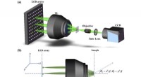
The new AET technique overcomes these challenges by using a high-energy electron beam to penetrate the material and create a 3D map of the atomic structure. The electron beam is focused to a very small spot, allowing the researchers to resolve individual atoms.
The researchers used AET to study a variety of amorphous materials, including glass, metal alloys, and semiconductors. They found that the atomic structure of these materials is not as random as previously thought. Instead, there are some degree of short-range order, or clustering, of atoms. This clustering can have a significant impact on the properties of amorphous materials, such as their strength, hardness, and electrical conductivity.
The new AET technique provides a powerful new tool for studying the structure of amorphous materials. This information could lead to the development of new materials with improved properties for a variety of applications.
The research was published in the journal Nature Materials.