Radiation damage is a serious problem in many fields, including medicine and materials science. It can cause significant degradation in the properties of materials, and it can also lead to harmful side effects in patients undergoing radiation therapy. Despite its importance, the exact mechanisms of radiation damage are still not fully understood, particularly for organic materials such as biological tissue and pharmaceuticals.
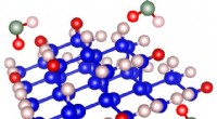
The new study, published in the journal Nature Physics, provides a major step forward in our understanding of radiation damage. The team used the X-ray free-electron laser LCLS at SLAC National Accelerator Laboratory in California to generate intense pulses of X-rays that were used to irradiate a crystal of the organic molecule tetraphenylcyclopentadienone (TPCP). The X-rays created damage to the crystal lattice, and the team used a variety of techniques to measure the damage in real time.
The results of the study show that radiation damage initiates through a process called "ionization-induced bond breaking." This occurs when an X-ray photon knocks an electron out of an atom or molecule, creating an unstable, highly reactive species called a "radical." The radical can then react with other molecules in the crystal, causing damage to the crystal lattice.
The team also observed that the damage was localized to the region of the crystal that was irradiated by the X-rays. This suggests that radiation damage can be minimized by using highly focused X-ray beams, which would allow researchers to study materials at the atomic level without causing significant damage.
The new study provides a detailed, atomic-level understanding of how radiation damage occurs in organic materials. This information is essential for developing new strategies to prevent or minimize radiation damage in a wide range of applications, including medicine, materials science, and X-ray imaging.
In addition to the researchers from DESY, MPSD, Aarhus University, and the University of Hamburg, the team also included researchers from UC Berkeley, the University of Chicago, and the University of California, Irvine.