Scientists have long known that antibiotics work by targeting the bacterial cell membrane, but the exact mechanism by which they do this has not been fully understood. The new images show that the antibiotics bind to the cell membrane and then form small, hole-like structures that grow and eventually burst the cell.
This process takes place on a very fast timescale, with the entire sequence of events occurring within milliseconds. The images were captured using a technique called X-ray free-electron laser (XFEL) microscopy, which allows scientists to capture images of objects moving at extremely high speeds.
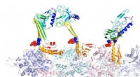
The researchers used this technique to image bacteria treated with a combination of two antibiotics, vancomycin and moenomycin A. Vancomycin is a antibiotic that has been used for decades to treat bacterial infections, while moenomycin A is a newer antibiotic that has been shown to be effective against some multi-drug resistant bacteria.
The images showed that the two antibiotics work together to kill bacteria. Vancomycin binds to the cell wall of the bacteria and inhibits the synthesis of new cell wall material, which weakens the cell. Moenomycin A binds to the cell membrane and causes the formation of holes in the membrane, which eventually leads to the destruction of the cell.
These findings could lead to the development of new antibiotics and treatments for bacterial infections. By targeting the bacterial cell wall, vancomycin and moenomycin A are able to kill bacteria that are resistant to other antibiotics. The combination of these two antibiotics could be used to treat infections that are caused by multi-drug resistant bacteria.
The researchers say that the new images provide a valuable tool for understanding how antibiotics work and could lead to the development of new antibiotics and treatments for bacterial infections.