By Kevin Beck | Updated Aug 30, 2022
Histology is the microscopic examination of tissues, extending the study of individual cells to the functional organization of cellular communities.
In clinical practice, rapid histological assessment can inform critical surgical decisions—such as whether to excise suspicious tissue during an operation—by revealing cancerous changes or other pathology.
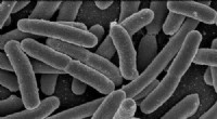
Microbiology underpins modern medicine, especially in diagnosing and controlling infectious diseases. Pathogens—bacteria, viruses, fungi, and protozoa—are too small to see unaided, yet they drive many illnesses.
Without microbiology, we would lack the knowledge to identify the causative agents of disease, differentiate between pathogen types, and develop targeted therapies.
The first compound microscope, a device using multiple lenses to magnify, appeared in 1590. While several inventors worked on such a device, the Jensen family—Hans and Zacharias—are credited with its creation.
It wasn’t until the 1660s that scientists began exploring microscopes for studying microscopic life. Soon after, Antony Van Leeuwenhoek observed the first bacteria, opening the field of microbiology.
Human tissues are broadly classified into four categories:
Each type derives from distinct embryonic layers and serves specialized functions.
Preparing histology slides requires specialized equipment beyond what a home laboratory offers. Thin sections are typically produced using a vibratome, a precision microtome that cuts slices suitable for microscopic analysis.
Additional essential instruments include:
Protocols vary between laboratories and depend on the sample type and diagnostic goal. Always adhere to institutional safety guidelines and verify that all reagents and equipment are handled correctly.