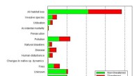
Here's a breakdown:
* Chromatin: The DNA-containing material within the nucleus, often stains darkly with basic dyes like hematoxylin.
* Nucleoli: Dense areas within the nucleus involved in ribosome synthesis, often stain darkly as well.
* Ribosomes: Sites of protein synthesis, can be stained with specific dyes that bind to their RNA content.
* Endoplasmic reticulum (ER): A network of membranes involved in protein synthesis and lipid metabolism. The rough ER, studded with ribosomes, often stains darker than the smooth ER.
* Golgi apparatus: A series of flattened sacs involved in processing and packaging proteins, can be stained with specific dyes that bind to its membranes.
* Mitochondria: Powerhouses of the cell, responsible for ATP production. They can be stained with dyes like Janus green B.
* Lysosomes: Sac-like organelles containing enzymes for cellular digestion, can be stained with specific dyes that bind to their enzymes.
* Cytoplasm: The jelly-like substance that fills the cell, can be stained with various dyes depending on the cellular components it contains.
Important Note: The specific staining properties of these organelles depend on the type of dye used and the chemical composition of the organelle.
Example: Hematoxylin, a basic dye, stains the acidic components of the nucleus (like DNA) blue. Eosin, an acidic dye, stains the basic components of the cytoplasm (like proteins) pink. This is why you often see a blue nucleus and pink cytoplasm in stained cells.