* Light Microscope (LM): While a basic light microscope can show the outline of some larger organelles, its resolution is limited. You'll need a compound light microscope with specialized techniques like:
* Phase Contrast Microscopy: Enhances the contrast of transparent structures, allowing you to see organelles like nuclei and vacuoles.
* Differential Interference Contrast (DIC) Microscopy: Creates a 3D-like image, useful for visualizing the internal structure of organelles.
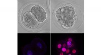
* Fluorescence Microscopy: Uses fluorescent dyes or proteins that bind to specific organelles, allowing you to see their location and sometimes their activity.
* Electron Microscope (EM): Offers much higher magnification and resolution than light microscopes, making them ideal for studying the fine details of organelles. There are two main types:
* Transmission Electron Microscope (TEM): A beam of electrons passes through the specimen, creating a detailed image of its internal structure. TEM is often used to study the internal organization of organelles like mitochondria, Golgi apparatus, and endoplasmic reticulum.
* Scanning Electron Microscope (SEM): A beam of electrons scans the surface of the specimen, creating a 3D image. SEM is useful for visualizing the external shape and surface features of organelles.
Ultimately, the best microscope for studying organelles depends on the specific research question and the desired level of detail.