Abstract:
Cell division is a fundamental biological process that ensures the growth, development, and reproduction of all living organisms. Understanding the intricate mechanisms underlying cell division is crucial for gaining insights into various cellular processes and diseases. However, the dynamic and complex nature of cell division poses significant challenges for traditional imaging techniques. Superresolution microscopy, with its ability to overcome the diffraction limit of light and provide nanoscale resolution, has emerged as a powerful tool for visualizing and studying cell division in unprecedented detail. This article explores the transformative capabilities of superresolution microscopy, highlighting how it enables researchers to zoom across time and space simultaneously, capturing the intricate details of cell division with exceptional precision and clarity. By delving into the realm of superresolution imaging, we gain a deeper understanding of the fundamental principles and advancements that have revolutionized the study of cell division.
Introduction:
Cell division is a tightly regulated process involving a series of precisely orchestrated events that lead to the duplication and segregation of genetic material into two daughter cells. This complex process encompasses various stages, including DNA replication, chromosome condensation, spindle formation, and cytokinesis. Traditional imaging techniques, such as widefield and confocal microscopy, have been extensively used to study cell division, but their limited resolution often impedes the visualization of fine cellular structures and dynamics.
Superresolution Microscopy: A Revolution in Imaging:
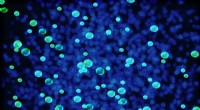
Superresolution microscopy represents a breakthrough in optical imaging, breaking the diffraction barrier that restricts the resolution of conventional microscopes. By employing advanced techniques such as stimulated emission depletion (STED), photoactivated localization microscopy (PALM), stochastic optical reconstruction microscopy (STORM), and structured illumination microscopy (SIM), superresolution microscopy enables the visualization of cellular structures and processes with nanoscale precision.
STED Microscopy:
STED microscopy utilizes a focused doughnut-shaped beam of light to selectively excite and inhibit fluorophores, allowing for targeted and high-resolution imaging. This technique has been instrumental in visualizing cellular structures such as microtubules, centrioles, and kinetochores, which play crucial roles in cell division.
PALM and STORM:
PALM and STORM are single-molecule localization techniques that enable the precise determination of the positions of individual fluorophores within a sample. By sequentially activating and imaging single molecules, these methods achieve superresolution images with exceptional detail. PALM and STORM have been extensively used to study dynamic cellular processes, including the assembly and disassembly of the mitotic spindle during cell division.
SIM Microscopy:
SIM microscopy employs structured illumination patterns to generate high-resolution images. By projecting a series of patterned light onto the sample and analyzing the resulting interference patterns, SIM microscopy achieves improved resolution compared to conventional wide-field microscopy. This technique has been utilized to study various aspects of cell division, including chromosome organization and cytokinesis.
Applications of Superresolution Microscopy in Studying Cell Division:
1. Visualization of Spindle Assembly and Dynamics:
Superresolution microscopy has provided unprecedented insights into the intricate details of spindle assembly and dynamics during cell division. Researchers have been able to visualize the organization of microtubules, the attachment of chromosomes to the spindle, and the forces generated during chromosome segregation.
2. Insights into Kinetochore Structure and Function:
Kinetochores, the protein complexes that connect chromosomes to the spindle, have been extensively studied using superresolution microscopy. This has led to a deeper understanding of their structure, composition, and interactions, shedding light on the mechanisms underlying chromosome segregation.
3. Cellular Membrane Dynamics:
Superresolution microscopy has also been instrumental in visualizing and understanding the dynamics of cellular membranes during cytokinesis, the process that separates the two daughter cells. Researchers have gained insights into the formation, constriction, and resolution of the contractile ring, elucidating the mechanisms involved in membrane remodeling and cell division completion.
Conclusion:
Superresolution microscopy has revolutionized the field of cell division research, empowering researchers to zoom across time and space simultaneously and capture the intricate details of this fundamental biological process with exceptional precision and clarity