One key aspect revealed by these experiments is the role of membrane dynamics during phagocytosis. As a cell encounters a solid particle, its plasma membrane undergoes significant remodeling. Specialized membrane structures, such as pseudopodia and phagocytic cups, extend and wrap around the particle, effectively engulfing it. These membrane extensions are driven by the actin cytoskeleton, a network of protein filaments that provides the cell with structural support and the ability to move.
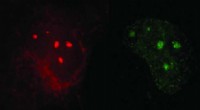
High-resolution imaging techniques, such as live-cell microscopy and super-resolution microscopy, have allowed scientists to visualize the intricate details of the phagocytic process. These techniques have captured the dynamic interactions between the cell membrane, the actin cytoskeleton, and various signaling molecules that orchestrate phagocytosis. By manipulating these cellular components through genetic or pharmacological interventions, researchers have gained a deeper understanding of the molecular mechanisms underlying phagocytosis.
Another important finding from these lab experiments is the involvement of specific receptors on the cell surface. Phagocytic cells, such as macrophages and neutrophils, express receptors that recognize and bind to specific molecules or ligands present on the surface of solid particles. This interaction triggers intracellular signaling cascades, leading to the activation of phagocytosis. The identity of these receptors and their ligands is crucial for the specific recognition and engulfment of different types of particles.
Furthermore, high-resolution experiments have revealed the existence of specialized intracellular compartments involved in phagocytosis. Once engulfed, the solid particles are enclosed within membrane-bound vesicles called phagosomes. These phagosomes then fuse with lysosomes, acidic organelles containing degradative enzymes. The acidic environment and enzymes within lysosomes break down the ingested particles into smaller components that can be recycled or utilized by the cell.
In summary, high-resolution lab experiments have greatly enhanced our understanding of the cellular machinery and molecular mechanisms involved in phagocytosis. By visualizing the dynamic processes and interactions at the nanoscale, scientists have gained insights into how cells recognize, engulf, and digest solid particles, contributing to our knowledge of fundamental cellular processes and immune responses.