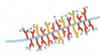
The researchers used a technique called X-ray crystallography, which allowed them to determine the three-dimensional structure of the protein complex. They found that the two proteins, called the coat protein and the movement protein, interact in a specific way to form a hexagonal "dimer of dimers" structure, which is the basic building block of the BMV capsid. This structure is characterized by two pairs of proteins arranged in a hexagonal shape.
This finding provides new insights into how viruses self-assemble and could potentially aid in the development of antiviral therapies. By understanding the molecular mechanisms underlying the formation of viral capsids, scientists can design drugs that target and disrupt the self-assembly process, preventing the virus from forming a protective shell and replicating.
"Our study provides a crucial piece of the puzzle in understanding how viruses assemble their capsids," said Eva-Maria Strasser, a postdoctoral researcher in the Department of Molecular Biology at UC Berkeley and lead author of the study. "This knowledge could lead to the development of new strategies to combat viral infections."
The researchers also gained insight into the role of the movement protein in the assembly process. The movement protein is known to be involved in the transport of the viral genetic material from the cell's nucleus to the site of capsid assembly, but its role in the actual assembly process was not well understood. The study revealed that the movement protein plays a structural role in the formation of the hexagonal building block, suggesting that it has dual functions in the viral life cycle.
"The movement protein seems to have two jobs: it helps the genetic material get to where it needs to go, and it also helps to build the capsid," said Jennifer Doudna, a Howard Hughes Medical Institute investigator, professor of molecular and cell biology, and senior author of the study.
The research team plans to further investigate the role of the movement protein in the assembly process and to explore how the findings can be applied to other viruses. They hope that this work will contribute to the development of new antiviral therapies and a deeper understanding of viral biology.