3PA has several potential advantages for bio-imaging over 1PA and 2PA. First, 3PA can provide deeper tissue penetration because the longer wavelength light used for 3PA is less scattered and absorbed by tissue components such as water and hemoglobin. Second, 3PA can be used to excite fluorescence in specific molecules with high selectivity because the excitation wavelength can be precisely tuned to match the absorption spectrum of the target molecule. Third, 3PA can generate higher resolution images because the smaller focal volume used for 3PA microscopy results in less photobleaching and photodamage to the sample.
Despite these potential advantages, 3PA is still not widely used for bio-imaging due to several challenges. First, the efficiency of 3PA is typically very low, requiring high laser powers that can damage biological samples. Second, the excitation wavelength for 3PA is often in the ultraviolet (UV) range, which can be harmful to cells. Third, the development of suitable 3PA probes is still in its early stages.
As these challenges are overcome, 3PA is likely to become a more important tool for bio-imaging. Its unique combination of deep tissue penetration, high selectivity, and high resolution make it ideal for a variety of applications, including in vivo imaging, fluorescence resonance energy transfer (FRET), and super-resolution microscopy.
Here are some specific examples of how 3PA has been used for bio-imaging:
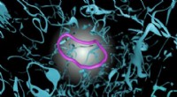
* 3PA microscopy has been used to image blood vessels in the brain of a living mouse. The 3PA signal was generated by a fluorescent dye that was specifically taken up by endothelial cells, the cells that line the blood vessels. This study demonstrated the potential of 3PA for in vivo imaging of deep tissue structures.
* 3PA FRET has been used to study protein interactions in live cells. In this technique, two different fluorescent dyes are attached to two different proteins of interest. When the proteins interact, the dyes come close together and the 3PA signal from one dye is transferred to the other dye. This allows researchers to monitor protein interactions in real time and with high spatial resolution.
* 3PA super-resolution microscopy has been used to image structures in cells with a resolution of less than 100 nanometers. This technique combines the high resolution of 3PA microscopy with the super-resolution capabilities of techniques such as stimulated emission depletion (STED) microscopy and photoactivated localization microscopy (PALM).
These examples demonstrate the potential of 3PA for bio-imaging. As the challenges associated with 3PA are overcome, this technique is likely to become increasingly important for a variety of applications in biomedical research and clinical diagnostics.