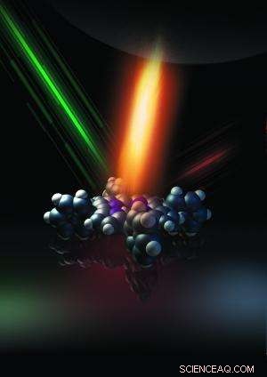
When a weak light beam of green color illuminates the molecule alone, the molecule is visible but lack of structural details (owing to the optical diffraction limit). However, when positioned under a tip, a much more intense and localized red-shifted light, produced by the plasmonic field, is acting on the molecule. The combination of both beams projects the vibrational fingerprints of the molecule into the emitting beam, chemically resolving the inner structure of the molecule with sub-nm resolution. Credit: Dong Xie and Rongting Zhou.
(Phys.org) —A team of researchers working at China's University of Science and Technology has succeeded in developing a chemical mapping technique capable of revealing the constituent atoms of a single molecule. In their paper published in the journal Nature, the team describes how they combined Raman spectroscopy with a scanning tunneling microscope (STM) to allow for chemical mapping of a molecule to a resolution of less than 1nm.
Raman spectroscopy is where chemists shine a laser on a small group of molecules and then measure the light as it's bounced back. The photons from the light source cause the molecules to vibrate and to interact with the bonds that hold molecules together causing a shift in their frequency—the scattering that results is unique for each type of molecule and thus allows for the method to be used as a means of identifying molecule types.
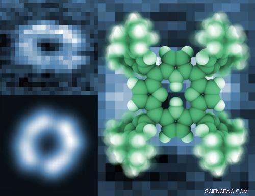
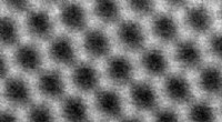
Top left: experimental map of an isolated porphyrin molecule for a given vibration frequency revealing the four-lobe pattern. Bottom left: theoretical calculation of the same molecular vibration showing its fingerprint. On the right: molecular structure of the porphyrin used in the experiment. Credit: Guoyan Wang and Yan Liang.
A STM is a device that allows for creating images of materials at the atomic level—one of its unique features is the very tiny metal tip used at the point of scanning. In this new effort the researchers combined Raman spectroscopy with STM to allow for unprecedented levels of molecular mapping.
Prior research has shown that when a STM tip is placed within nanometers of certain metals, plasmonic excitation occurs that when combined with Raman scattering can allow for mapping molecules to within 10nm. In this new research, the team has found that if the frequency of the plasmonic excitation is adjusted to match the molecular vibrations caused by photons from the laser light, the Raman signal is increased sharply, resulting in an ability to map the molecule being studied to less than 1nm.
Owing to the optical diffraction limit, a single porphyrin molecule cannot be resolved by conventional optical imaging with a green laser alone. However, when the molecule is positioned under a tip, a much more intense and localized red-shifted light, produced by the plasmonic field, is acting on the molecule. The combination of both beams projects the vibrational fingerprints of the molecule into the emitting beam, chemically resolving the inner structure of the molecule with sub-nm resolution.
The researchers note their technique is still in the very early stages of development—thus far they've only been able to use it on one molecule—a ring-shaped porphyrin. The process they note, is difficult and can take weeks or months carry out making its application impractical at this point for general research efforts. Also it only works when the molecule under study is held in a vacuum and in a -200° C environment. If the technique can be fined tuned however, it will allow future chemists to identify the atoms in individual molecules. Such a tool could open the door to new ways to study molecules at the nano-scale level as well as the bonds that hold them together.
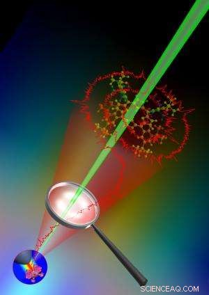
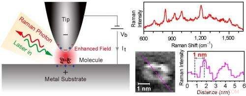
Left: Schematic diagram of tunneling-controlled tip-enhanced Raman scattering (TERS) in a confocal-type side-illumination configuration, in which Vb is the sample bias and It is the tunneling current. A laser light is focused into the nanocavity defined by the scanning tunneling microscope (STM) tip and substrate. The strong local plasmonic field generated by the incident laser causes the enhancement of Raman scattering from the single molecule underneath the tip. Top right: TERS spectrum acquired on the lobe; Bottom right: TERS map for the vibrational mode at about 817 cm-1 and corresponding line profile. Credit: Zhenchao Dong
© 2013 Phys.org