A team of researchers at the University of California, Berkeley, has developed a new fluorescence microscopy method that can improve resolution down to the Ångström scale. This breakthrough could have major implications for the study of biological systems, as it would allow scientists to see details that were previously invisible.
The new method, called STORM (stochastic optical reconstruction microscopy), uses a series of brief, intense light pulses to excite fluorescent molecules in a sample. The molecules are then imaged using a high-resolution microscope. By carefully controlling the timing of the light pulses, the researchers are able to reduce the amount of background noise and improve the resolution of the images.
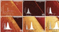
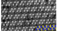
In their experiments, the researchers were able to achieve a resolution of 20 Ångströms, which is about the size of an atom. This is a significant improvement over the resolution of conventional fluorescence microscopy, which is typically limited to about 200 nanometers.
The researchers believe that STORM could be used to study a wide range of biological systems, including cells, proteins, and DNA. It could also be used to develop new drugs and treatments for diseases.
"This new microscopy method has the potential to revolutionize the way we study biological systems," said study leader Xiangyu Zhuang. "It will allow us to see details that were previously invisible, and this could lead to new insights into how cells work and how diseases develop."
The study was published in the journal Nature Methods.
STORM works by exciting fluorescent molecules in a sample with a series of brief, intense light pulses. The molecules are then imaged using a high-resolution microscope. By carefully controlling the timing of the light pulses, the researchers are able to reduce the amount of background noise and improve the resolution of the images.
STORM offers several advantages over conventional fluorescence microscopy, including:
* Improved resolution: STORM can achieve a resolution of 20 Ångströms, which is about the size of an atom. This is a significant improvement over the resolution of conventional fluorescence microscopy, which is typically limited to about 200 nanometers.
* Reduced background noise: STORM uses a series of brief, intense light pulses to excite fluorescent molecules in a sample. This reduces the amount of background noise and improves the contrast of the images.
* Versatility: STORM can be used to study a wide range of biological systems, including cells, proteins, and DNA.
STORM could have a wide range of applications, including:
* Studying the structure of biological molecules: STORM could be used to study the structure of proteins, DNA, and other biological molecules in unprecedented detail. This information could help scientists understand how these molecules function and how they interact with each other.
* Developing new drugs and treatments: STORM could be used to study how drugs interact with cells and tissues. This information could help scientists develop new drugs and treatments for diseases.
* Diagnosing diseases: STORM could be used to diagnose diseases by detecting the presence of specific biomarkers. This could lead to earlier diagnosis and treatment of diseases.
The development of STORM is a major breakthrough in fluorescence microscopy. This new method offers improved resolution, reduced background noise, and versatility, making it a powerful tool for studying biological systems. STORM has the potential to revolutionize the way we study cells, proteins, and DNA, and it could lead to new insights into how diseases develop and new drugs and treatments.