Here's how it works:
* Electron Beam: An SEM uses a focused beam of electrons to scan the surface of a specimen.
* Interaction: The electrons interact with the atoms on the surface, causing various signals to be emitted, such as:
* Secondary Electrons: These are emitted from the surface of the specimen due to the energy transferred from the primary electron beam. They provide information about the specimen's topography (surface features).
* Backscattered Electrons: These are primary electrons that have been deflected back from the specimen. They provide information about the specimen's composition, as different elements scatter electrons differently.
* Image Formation: The emitted signals are detected and used to create an image of the specimen's surface.
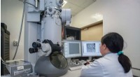
SEMs provide detailed images of the surface of a specimen, revealing its three-dimensional structure and composition. They are used in a wide range of fields, including materials science, biology, and medicine.