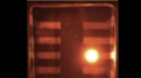
The image formed by an electron microscope is typically sharp at the center and blurred at the edges due to several factors:
1. Spherical Aberration:
Spherical aberration is an inherent limitation of electron lenses caused by the focusing of electrons in a non-perfectly spherical field. This leads to distortions and blurring of the image, particularly at the edges.
2. Electron Scattering:
As the electron beam passes through the specimen, it interacts with the atoms and molecules present. The heavier the atoms, the more they scatter electrons. This scattering effect is more pronounced towards the edges of the sample where the electrons travel through a greater amount of material, leading to a loss of resolution and a blurred appearance.
3. Edge Effects:
At the edges of the specimen, the electron beam encounters abrupt changes in the material's thickness or density. This can cause diffraction and scattering of electrons, resulting in edge effects that contribute to image blurriness.
4. Specimen Preparation:
Sample preparation for electron microscopy involves thin sectioning or coating the specimen, which can introduce artifacts or damage the edges of the sample. These preparation-related factors can also contribute to image blurring at the edges.
5. Imperfect Focusing:
Precise focusing is crucial in electron microscopy. If the microscope is not perfectly focused, the image may appear blurred, especially at the edges.
To minimize these effects and obtain sharp images, electron microscopes are equipped with advanced correction systems such as spherical aberration correctors and image processing algorithms to enhance the quality and resolution of the images.