The LCLS team, which includes scientists from the New York University School of Medicine, the University of Wisconsin-Milwaukee and Argonne National Laboratory, also managed to record the first X-ray laser scattering images of an intact virus, vaccinia virus, which is about the size of the smallest bacteria.
The results, reported in two papers in Nature Communications, demonstrate the X-ray laser's promise as a powerful new tool for exploring biological structures.
"This was the first time we were able to use X-ray lasers to image these two very important classes of biological samples, which hold valuable information that could lead to new ways to treat diseases," said SLAC staff scientist Henrik Lemke, who is the corresponding author of the study on protein crystals.
To get the job done, the team had to make a few adjustments to the hard X-ray beam at SLAC's Linac Coherent Light Source (LCLS), which provides ultrabright, ultrashort pulses of X-rays.
One challenge was that the X-ray pulses were too bright and concentrated, threatening to damage or destroy the delicate samples—and the surrounding sample holder.
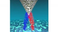
"Our beam is normally about the size of a very thin human hair, but we made the beam a hundred times larger so we could scatter and diffract the X-rays more gently off of the samples," said LCLS instrument scientist and study co-author Schuyler Brown.
Researchers also needed to develop new sample preparation techniques to prevent damage caused by the intense X-ray beam. Because the laser's flashes last only femtoseconds (quadrillionths of a second), damage occurs within just ten-quadrillionths of a second.
Using a technique known as serial femtosecond crystallography, the scientists fired intense X-ray pulses one at a time at thousands of tiny crystals to create a wealth of diffraction patterns—patterns of scattered X-rays that contain structural information about the crystals.
"In most cases, we only fired one X-ray pulse at each crystal because the first flash would destroy it," said study co-author Thomas White of New York University School of Medicine. "As a result, each flash generated only one diffraction pattern. Then we combined all the patterns to reconstruct a three-dimensional image of the crystals' structures."
With this technique, the team resolved the structure of protein crystals known as photosystem II, which are responsible for converting sunlight into chemical energy during photosynthesis. The results represent the smallest photosystem II structure yet obtained.
The team's scattering images of vaccinia virus also produced some surprises, showing that some of the viruses in the sample were in an unexpected, highly symmetrical conformation. This type of conformation could affect how the virus interacts with hosts and might reveal an Achilles' heel that could be targeted by antiviral drugs.
"This is another great example of how the X-ray laser enables researchers to see things in biology they've never seen before," said SLAC Director Mike Witherell. "By peering into the details of viruses or proteins that aren't visible with any other technique, we're not only gaining a deeper understanding of the natural world, but opening the door to new ways to fight disease and create renewable energy."
SLAC's LCLS is scheduled for an upgrade in 2018, which will dramatically increase its power, opening up even more biological imaging possibilities. Future instruments at SLAC's future X-ray laser, LCLS-II, will also support biological imaging.
The research was funded by the Department of Energy's Office of Science, the National Institutes of Health, the University of Wisconsin-Milwaukee and New York University School of Medicine.