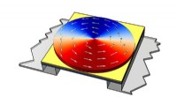
The imaging device, called the "Digital Holographic Microscopy (DHM) system," uses coherent light to capture three-dimensional information about a swimming fish. The system consists of a high-resolution camera and a laser, which illuminates the fish tank from below. As the light passes through the water and interacts with the fish, it creates a distorted wavefront. This wavefront is then captured by the camera, which records the interference patterns created by the interaction of the light and the fish.
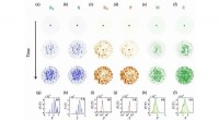
By analyzing the recorded interference patterns, the researchers can reconstruct a three-dimensional image of the fish's body and its movements. This allows them to track the fish's body kinematics and calculate the forces generated by different fins. The DHM system provides much higher spatial and temporal resolution compared to traditional imaging techniques, allowing for detailed analysis of fish swimming biomechanics.
The researchers used the DHM system to study the swimming behavior of zebrafish, a small freshwater fish commonly used in biological research. They found that the pectoral and pelvic fins play a significant role in generating thrust for forward propulsion, while the caudal fin (tail) contributes mainly to maneuvering and stability. The study also revealed that the zebrafish can modulate the force production and movement of its fins to achieve different swimming speeds and turn angles.
The findings from this research have implications for understanding fish locomotion, ecological interactions, and the evolution of swimming adaptations. The DHM system provides a powerful tool for studying the biomechanics of fish swimming and opens new avenues for exploring the complex underwater world of fish.