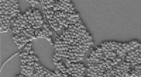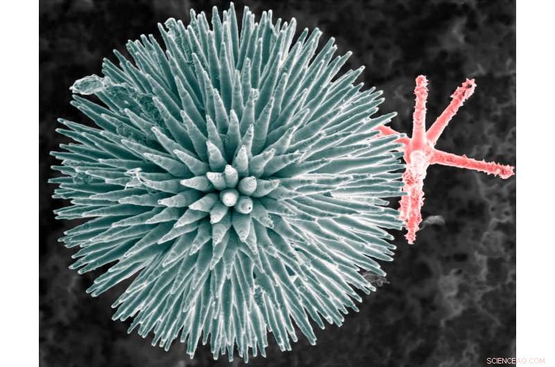
Electron microscopy image of glass spicules from the sponge Geodia cydonium. Credit: Zlotnikov Group, B CUBE, TU Dresden
A team of researchers with members from France, Germany and Israel has found that proteins that make up axial filaments are responsible for the means by which sea sponges develop glass skeletons. In their paper published on the open access site Science Advances, the group describes their study of the sea creatures, what they found and why they believe their work could lead to advances in the creation of materials for use in new opto-electronic devices.
Sea sponges, the researchers note, are some of the oldest creatures on Earth—fossil records show they date back a half-billion years. Over that time, the researchers also note, they have evolved to grow spiky glass structures that make up their unique appendages (oddly, they have no tissue or organs). They further note that little research has been done to better understand how such structures emerge as the creature matures, which is unfortunate, because it is clear they do so without the need for fiery hot furnaces. To learn how the sea creatures are able to create glass structures, the researchers looked at three types of the sponges and more specifically at their distinct spicules (needle-shaped structures).
The team used X-ray diffraction and an electron microscope to take a closer look at the spicules and the axial filaments around which they form. In so doing, the group found that the filaments were made from proteins stacked in a hexagonal crystalline structure. The researchers noted that the structures were nearly the same for all three of the sponges they looked at, though each have unique spicule shapes: needle-like for Thethyra aurantium, three-way branches for Stryphnus ponderosus and spikey orbs for Geodia cydonium. The differences in the resulting shapes, the team found, were due to how the proteins were spaced and arranged. The glass exists as deposits of silica on the spicula—the protein serves as a template.
The researchers suggest more study of the creatures may lead to the development of a similar mechanism for the manufacture of tiny glass components for use in opto-electronic devices, plasmonics and perhaps solar cells.
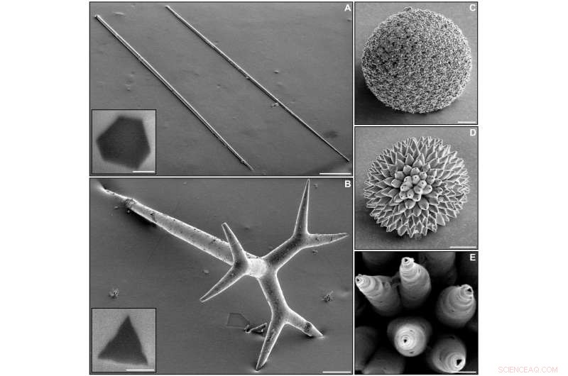
The morphology of demosponge spicules and their inner axial filaments as revealed by scanning electron microscopy. (A) Megascleres from the demosponge T. aurantium. Scale bar, 100 mm. Inset: Cross section of the spicule obtained by focused ion beam (FIB). Scale bar, 1 mm. (B) Megasclere from the demosponge S. ponderosus. Scale bar, 100 mm. Inset: Cross sections of the main shaft of the spicule obtained by FIB. Scale bar, 1 mm. Note: Some spicule tips were broken during sample preparation and appear to be flat. (C to E) Microscleres from the demosponge G. cydonium at different maturation levels, from a fully mature spicule to an immature one, respectively. Credit: Schoeppler et al., Sci. Adv. 2017;3: eaao2047
© 2017 Phys.org