By Pete Collins | Updated Aug 30, 2022
Electrophoresis separates macromolecules such as proteins and nucleic acids by size, charge, and other physicochemical properties. In charge‑based separations, a buffer solution transmits the electrical field and maintains a stable pH, preserving the native charge and structure of the analytes. This stability is critical for accurate resolution.
Electrophoresis leverages an applied electric field (or a chemical gradient in specialized techniques) to move charged molecules through a gel matrix. Molecules migrate toward the electrode of opposite charge: negatively charged species travel to the anode, positively charged species to the cathode. Because larger molecules experience more friction within the gel, they move more slowly than smaller ones, allowing size‑based separation. The distance migrated can be plotted on a logarithmic scale to estimate molecular weight or fragment length.
DGGE introduces a denaturant gradient—typically a mixture of urea and formamide—within the gel. As DNA fragments traverse the gradient, increasing denaturant concentrations progressively destabilize the double helix. Each fragment stops migrating once the local denaturant concentration reaches its melting point. This technique exploits sequence‑dependent melting behavior to resolve DNA fragments of identical length but different sequence.
In charge‑based electrophoresis, the buffer’s ionic species conduct the applied electric field through the gel, ensuring uniform current distribution. Simultaneously, the buffer’s weak acid–base pair maintains the pH within a narrow window. Because the charge state and three‑dimensional structure of proteins and nucleic acids are pH‑dependent, a stable pH prevents unintended conformational changes that could compromise separation fidelity.
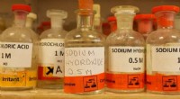
Choosing the right buffer hinges on the desired pH range and the need to minimize ionic strength, which could otherwise generate excessive heat and distortion. Commonly used buffers include:
For optimal performance, the buffer’s pKa should be close to the target pH, and the overall ionic strength should be low enough to limit current‑induced heating while still allowing efficient charge transfer.
By carefully selecting and maintaining buffer conditions, researchers can achieve reproducible, high‑resolution separations essential for downstream analyses.