Electron microscopes use a beam of electrons instead of light to illuminate a sample. Here's a breakdown of how it works:
1. Electron Gun: This part generates a stream of electrons. It's similar to a cathode ray tube found in old TVs.
2. Electromagnetic Lenses: These lenses are not made of glass but use electromagnetic fields to focus the electron beam. They act like optical lenses, bending the electron paths to create a sharp image.
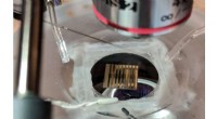
3. Specimen: The sample you want to examine is placed in the microscope's vacuum chamber. This vacuum is crucial because electrons are easily scattered by air molecules.
4. Interaction: The electron beam interacts with the specimen in various ways, depending on the type of microscope:
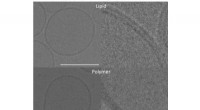
* Transmission Electron Microscopy (TEM): Electrons pass through the sample. Thinner areas allow more electrons to pass, creating a darker image. This technique is good for studying the internal structure of cells and materials.
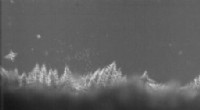
* Scanning Electron Microscopy (SEM): Electrons are scanned across the surface of the sample. The interaction between the electrons and the sample generates signals that are used to create a 3D image of the surface. This technique is great for visualizing the surface features of objects.
5. Detectors: The detectors capture the signal from the electron beam after its interaction with the sample. This signal is then processed to generate an image.
6. Image Formation: The image is formed based on the intensity of the detected signal. In TEM, a brighter area means more electrons passed through, indicating a thinner part of the sample. In SEM, a brighter area indicates a higher number of electrons emitted from that point.
Key Advantages of Electron Microscopes:
* Higher Resolution: Electron microscopes can achieve much higher resolution than light microscopes, allowing scientists to see incredibly small details, even individual atoms.
* Versatile Applications: They are used in various fields, including biology, materials science, nanotechnology, and forensic science.
Key Limitations:
* Sample Preparation: Samples need to be thin enough for TEM or conductive for SEM, which can be complex and time-consuming.
* Vacuum Requirement: The need for a vacuum environment limits the study of living specimens.
In short, electron microscopes are powerful tools that use a beam of electrons to create high-resolution images of samples, allowing scientists to explore the microscopic world in unprecedented detail.