1. X-rays:
* Wavelength: 0.01 to 10 nanometers
* Pros: High energy and short wavelength allow them to penetrate matter and interact with electron clouds around atoms.
* Cons: High energy can damage molecules. Diffraction patterns are complex and require specialized techniques like X-ray crystallography to interpret.
2. Extreme Ultraviolet (EUV) Radiation:
* Wavelength: 1 to 121 nanometers
* Pros: Short wavelength suitable for imaging individual molecules.
* Cons: Requires specialized equipment and can damage samples. Used in high-resolution microscopy techniques like photoemission electron microscopy (PEEM).
3. Electron Microscopy:
* Not electromagnetic radiation: Uses a beam of electrons instead of light.
* Pros: Very high resolution, capable of imaging individual atoms and molecules.
* Cons: Requires special sample preparation and high vacuum conditions. Not suitable for live samples.
4. Scanning Tunneling Microscopy (STM):
* Not electromagnetic radiation: Uses a sharp tip to probe the surface of a material.
* Pros: Atomic resolution, can be used to image and manipulate individual molecules.
* Cons: Only works on conductive or semi-conductive materials, and requires high vacuum conditions.
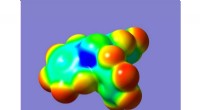
5. Atomic Force Microscopy (AFM):
* Not electromagnetic radiation: Uses a sharp tip attached to a cantilever to scan the surface of a material.
* Pros: High resolution, can be used to image biological samples, and can be used in liquid environments.
* Cons: Not as high resolution as STM, can be difficult to interpret complex structures.
In Summary:
While no single method can perfectly "see" molecules in all scenarios, a combination of these techniques provides a powerful toolbox for studying molecular structure and function. The choice of method depends on the specific application and the desired level of detail.