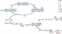
Mitosis:
1. Prophase: Chromosomes condense and become visible. Each chromosome consists of two identical sister chromatids held together at the centromere.
2. Metaphase: The chromosomes line up at the center of the cell (the metaphase plate). The spindle fibers, which are made of microtubules, attach to the centromeres of each chromosome.
3. Anaphase: The centromeres divide, and the sister chromatids are pulled apart by the spindle fibers. Each chromatid now becomes a separate chromosome, migrating to opposite poles of the cell.
4. Telophase: The chromosomes reach the poles and begin to decondense. The nuclear envelope reforms around each set of chromosomes.
Meiosis II:
1. Prophase II: The chromosomes condense again. They are already duplicated from meiosis I, so each chromosome consists of two sister chromatids.
2. Metaphase II: The chromosomes line up along the metaphase plate, with spindle fibers attaching to the centromeres.
3. Anaphase II: The centromeres divide, and the sister chromatids separate. They are now considered individual chromosomes and move towards opposite poles of the cell.
4. Telophase II: The chromosomes reach the poles, decondense, and nuclear envelopes form around them. The cell then divides (cytokinesis), resulting in four haploid daughter cells.
Key Points:
* Spindle fibers are crucial for separating the chromatids. They attach to the centromeres and pull the chromatids apart.
* Centromere division is essential for the separation of sister chromatids.
* The separation of chromatids ensures that each daughter cell receives a complete set of chromosomes.
In summary, the process of separating chromatids is a highly organized and regulated event that ensures accurate chromosome distribution during cell division.