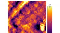
1. Atomic Force Microscopy (AFM):
- AFM is a technique that uses a sharp probe to scan the surface of a material. By scanning the probe over individual atoms, scientists can create three-dimensional images of the atomic structure on a surface.
2. Scanning Tunneling Microscopy (STM):
- STM is another scanning probe microscopy technique that uses a sharp conducting tip to scan a surface. When a voltage is applied between the tip and the material, a tunneling current flows between them. Variations in this tunneling current provide information about the electronic structure and topography of the atoms on the surface.
3. Transmission Electron Microscopy (TEM):
- TEM uses a focused beam of high-energy electrons to pass through a thin sample. The transmitted electrons interact with the atoms within the sample, producing images with high resolution. TEM can reveal the detailed internal structure of atoms and their arrangements in a material.
4. Scanning Electron Microscopy (SEM):
- SEM uses a focused beam of electrons to scan the surface of a material. The incident electrons interact with the sample's atoms, emitting secondary electrons and other signals that can be detected and used to create images of the surface topography and composition.
5. X-ray Crystallography:
- X-ray crystallography utilizes the scattering of X-rays by atoms within a crystal lattice to determine the arrangement and positions of atoms in a crystal. By analyzing the diffraction patterns produced, scientists can deduce the atomic structure and crystallographic properties of materials.
6. Molecular Modeling and Simulation:
- Scientists use computational techniques and software to create virtual models of molecules and atoms. These models can be used to simulate and visualize atomic interactions, behaviors, and properties at the molecular level.
7. Cryogenic Electron Microscopy (Cryo-EM):
- Cryo-EM is a specialized TEM technique performed at extremely low temperatures to prevent damage to biological samples. By rapidly cooling and preserving the sample in a vitreous ice, scientists can capture detailed images of individual molecules and macromolecular complexes, revealing their structures at near-atomic resolution.
These techniques and methods allow scientists to visualize atoms in various ways, providing valuable information about their structure, arrangement, and behavior within different materials and systems. The choice of visualization approach depends on the specific materials, properties, and level of detail required for the research investigation.