When scientists talk about crystals, they often mean single crystals. These highly ordered structures consist of atoms, molecules or ions arranged in a repetitive, three-dimensional pattern. Because their repeating building-block units are regular and stack neatly atop one another, single crystals tend to be strong, uniform and easy to characterize.
But nature seldom provides perfect single crystals. Instead, materials often come as polycrystalline aggregates, a hodgepodge of smaller, randomly oriented single crystals.
That disparity matters because the properties of a material strongly depend on how its atoms or molecules pack together. For instance, the performance of silicon solar cells and LEDs depends on the size and orientation of the material’s tiny single crystals.
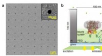
Now researchers reporting in the journal ACS Nano describe how they filmed the growth of crystals. The team, led by Yassar Dahman of the University of Virginia, used a microscopy method known as atomic force microscopy to watch how tiny crystals nucleate on a silicon substrate.
Atomic force microscopes use a sharp cantilever, similar to that of a scanning probe microscope, to scan the surface. As the cantilever moves across a sample, its vertical position is adjusted as needed to maintain a constant force between the tip and the surface. The resulting data can then be used to determine how the surface topography varies along the scan.
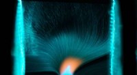
The group set up their instrument to scan an area slightly larger than 2 micrometers on a side, every 2 milliseconds — a process they continued for more than half an hour. The researchers’ video shows how nanometer-scale crystalline islands form on the substrate. The video also reveals that the islands grow quickly, merge with one another and move around the surface as the material rearranges itself, eventually forming larger and more perfect crystals.
“You can see a small island nucleate, and it will start growing and eventually hit another island and merge with it,” says Dahman.
Dahman notes that the film’s time scale is orders of magnitude faster than that of other techniques used to image the movement of atoms on surfaces, such as scanning tunneling microscopy. “What we’re showing here is very different from what we see with those techniques, which show static images because they’re probing the surface very slowly,” he says. “We’re seeing a movie, instead of a still image.”
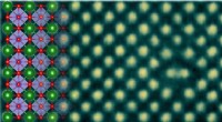
The technique also reveals that the islands initially have different structures, but then the most stable structure takes over as the crystals grow larger, Dahman says. “The more stable structure is the one with the lower surface energy,” he explains.
Dahman says that the team hopes to use the new microscopy method to study how different materials grow in real time, to learn more about why materials adopt specific crystal structures and to design better materials for various applications.
Matthew J. Highland of the University of Chicago, who was not involved in the research, says the work is “very intriguing” and “exciting.”
“The ability to observe the evolution of crystal growth in situ at the nanoscale is of great value to the field,” he says. And although the researchers imaged crystals growing on silicon, Highland notes that “this technique is equally applicable to a variety of other materials systems, including organic semiconductors, metal oxides and even biomolecules.”