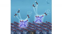
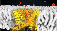
At these ultrafast timescales, the team observed the drugs creating microscopic pores in the bacteria's protective outer layer, causing its contents to leak out and the cell to die.
The experiments are some of the first of their kind and the results could pave the way for designing more effective antibiotics that target the bacteria membrane more specifically.
The research is published in the journal ACS Central Science.
How do antibiotics work?
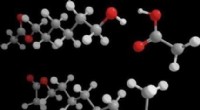
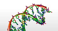
One way that antibiotics kill bacteria is by targeting and damaging the integrity of the bacterial outer membrane, composed of a phospholipid bilayer.
These are two layers of fat molecules (phospholipids) that form a barrier around all bacterial cells. The bilayer is a dynamic and complex structure that has unique physical properties, such as its fluidity, that depend on the types and number of lipids in the membrane.
Membrane disruption is a leading cause of bacterial death, but how exactly this happens has not been fully understood, largely because these events occur on exceptionally short timescales.
The current study addresses this knowledge gap by combining experiments with computational simulations to probe the initial processes involved in membrane damage.
Capturing the unseen
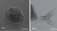
A team led by researchers from Oxford University and the University of California, San Diego, used high-speed atomic force microscopy (HS-AFM) to watch a membrane-disrupting antibiotic interacting with a bacterial membrane in near-real-time.
The high-speed instrument records the interactions between an oscillating nanoscale tip and a soft material at a rate of nearly 770,000 frames per second, revealing details that can't be seen using conventional imaging methods.
The team used the instrument's unique capabilities to capture the events that happen in the microseconds to milliseconds timescales after an antibiotic makes contact with the bacterial membrane, allowing them to observe the very beginning of the cell death process.
Professor Aleksander Bublitz from Oxford's Department of Chemistry, said: 'It's incredibly difficult to image fast processes at such small length scales, but by combining this tool with computational simulations we can begin to understand how antibiotic drugs cause membranes to break down. We can then use that information to design better drugs that specifically target the bacterial membranes and disrupt them more effectively.'
New insights
The research reveals, for the first time, the crucial role that the bacterial membrane fluidity and the antibiotic molecule binding dynamics play in the pore-forming process.
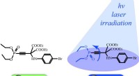
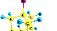
By mimicking the system with computer simulations, the researchers were able to see how the antibiotic works at the atomic level.
The simulations allowed them to identify particular membrane lipids and specific antibiotic-membrane interactions that are critical to the pore formation process.
This information can be used to develop more effective drugs by engineering specific molecules that target the identified lipids or antibiotic binding sites.