Photodisc/Photodisc/Getty Images
Prior to cell division, the DNA within the nucleus must be faithfully duplicated, meticulously inspected for errors, and organized into compact, chromatid-like structures. This intricate sequence ensures that each daughter cell receives an accurate copy of the genome.
Cell division, or mitosis, is a key component of the cell cycle. It follows a preparatory phase called interphase and a division phase known as the M phase. The M phase is subdivided into mitosis and cytokinesis—the latter being the physical split that yields two daughter cells. The classic stages of mitosis are prophase, metaphase, anaphase, and telophase, each contributing to the formation of identical daughter nuclei.
Interphase itself is divided into three substages: G1, S, and G2. During G1 (the first gap), the cell grows and synthesizes proteins. In S (synthesis) phase, DNA replication occurs, producing sister chromatids. G2 (the second gap) is dedicated to organelle duplication and a thorough review of the newly synthesized DNA for potential errors before the cell commits to division.
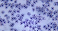
After replication in the S phase, each chromosome consists of two identical sister chromatids. In humans, the result is two complete sets of 46 chromosomes—23 from each parent. Unlike meiosis, mitosis does not involve the pairing of homologous chromosomes.
During the S phase, the cell duplicates its entire genome in a highly coordinated process that temporarily unwinds and exposes the DNA strands. This necessary decondensation increases the risk of breaks, so the cell expends significant energy and employs robust replication machinery to safeguard fidelity.
Once duplication is complete, the newly formed sister chromatids are compacted into short, thick, chromatid structures—essentially X‑shaped chromosomes. DNA does not exist in isolation; it is wrapped around histone proteins, forming chromatin. This condensation into tightly wound, cylindrical bundles strengthens the DNA and protects it from damage during the mechanical forces of mitosis.
Each condensed chromosome features a centromere—a specialized region that serves as the attachment point for spindle microtubules, enabling precise segregation of chromatids during cell division.
Before mitosis can proceed, the cell conducts a comprehensive inspection of the replicated DNA during the G2 phase. Dedicated DNA‑damage‑response proteins scan for nicks, breaks, or mismatches. If defects are detected, the checkpoint machinery halts progression, allowing repair processes to correct the issues. Only after passing the G2‑M checkpoint does the cell advance into mitosis.