Understanding the primary cells and tissues of the body is a cornerstone of biology education. Whether you study general biology, anatomy, or physiology, epithelial tissue will appear in every curriculum.
Why? Epithelial tissue is the most abundant of the four major tissue types—connective, muscular, nervous, and epithelial. It lines every organ and surface in the body, forming a protective and functional barrier.
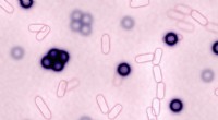
In histology, epithelial tissue is divided into two principal categories: stratified epithelium (multiple cell layers) and simple epithelium (single cell layer). This article focuses on the latter, exploring its basic architecture and the four distinct cell shapes.
Simple epithelial tissue consists of a single cell layer adhered to a connective‑tissue foundation known as the basement membrane. The cells are polarized, featuring a basal surface that contacts the basement membrane, an apical surface that faces the body’s lumen or external environment, and lateral borders that form strong intercellular junctions.
Although the overall organization is uniform, the shape of the cells within the single layer determines the tissue’s specific function and location. There are four primary types of simple epithelial tissue.
Squamous cells are flattened, creating the thinnest epithelial layer. Their narrow, elongated nuclei sit centrally. This minimal thickness sacrifices protection but excels in diffusion and exchange.
Key examples:
Cube‑shaped cells are slightly thicker than squamous cells, offering modest protection while retaining permeability. Each cell contains a centrally positioned, round nucleus.
Functions include secretion and absorption. Common sites:
Columnar cells are tall, column‑like, providing the greatest protection among simple epithelia. Their nuclei are typically located at the basal or lateral edges.
Two variants:
Although it is a single cell layer, pseudostratified epithelium displays nuclei at varying heights, creating a stratified appearance. It is usually ciliated.
Locations: