© rob_lan/iStock/GettyImages
The kingdom Protista is a diverse assemblage of microscopic eukaryotes that do not fit neatly into the kingdoms of plants, animals, or fungi. All protists possess a true nucleus and membrane‑bound organelles such as mitochondria and Golgi complexes. Modern phylogenomic studies have reorganized this group into several supergroups, linking many protists to other major lineages of life.
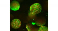
Volvox globator is a striking green alga that forms large, hollow, spherical colonies. Each colony consists of thousands of tiny flagellated cells that beat their whip‑like flagella to propel the entire sphere through water. Colonies can reach up to 2 mm (0.08 in) in diameter, making them visible to the naked eye. Reproduction occurs both sexually, with distinct male and female colonies, and asexually, when daughter colonies develop inside a parent colony.
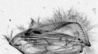
Common in freshwater habitats, Paramecium caudatum is a single‑cell organism roughly the size of a period (about 0.2–0.3 mm). Its surface is covered with densely packed cilia that beat rhythmically to propel the cell and to direct bacteria and other food particles into its oral groove. Inside the cell, food is stored in a vacuole for digestion, while waste is expelled through the cytostome. The organism contains two distinct nuclei: a large macronucleus that governs everyday cellular functions, and a smaller micronucleus involved in conjugation, a form of genetic exchange.
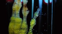
Physarum polycephalum is a true (plasmodial) slime mold. Visible colonies are yellow‑ish and irregular, with bulbous protrusions. The organism is a multinucleate mass of cytoplasm that forms when individual flagellated cells fuse. Although once grouped with fungi due to similar spore‑forming strategies, genetic analyses place it firmly within the Amoebozoa. Slime molds can also undergo a primitive sexual phase, exchanging genetic material across the plasmodium.
Most pond water contains a rich community of protists. To observe them, simply collect a drop of water in a syringe, hold the syringe upside down, and let a droplet hang from the tip. Direct a laser pointer through the droplet; the droplet will act as a lens, projecting a magnified image of the protists onto a nearby wall in a darkened room. For a more detailed view, a standard light microscope will reveal the structure and behavior of these microscopic life forms.