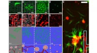
* Confocal microscopy uses a laser beam to illuminate a single point in the specimen at a time.
* This focused illumination eliminates out-of-focus light from other parts of the specimen, resulting in sharper images with increased contrast.
* By scanning the laser beam across the specimen, multiple images are captured at different depths.
* These images are then computationally combined to create a 3D reconstruction of the organism.
Other tools used in life science for microscopy include:
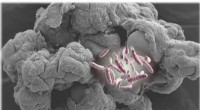
* Scanning Electron Microscope (SEM): Provides high-resolution images of the surface of a specimen, but only in 2D.
* Transmission Electron Microscope (TEM): Provides detailed images of the internal structure of a specimen, also in 2D.
* Light Microscopy: Uses visible light to illuminate the specimen, offering a less detailed view but with the advantage of being able to observe live organisms.
However, for obtaining detailed 3D images of microscopic organisms, confocal microscopy is the preferred choice.