1. Microscopy:
* Light Microscopy (LM): This is the most basic form of microscopy. It uses visible light to illuminate and magnify the sample.
* Bright-field microscopy: The most common type, it produces an image where the specimen is dark against a bright background.
* Phase-contrast microscopy: This technique enhances the contrast of transparent specimens by exploiting differences in refractive index.
* Differential interference contrast (DIC) microscopy: This technique uses polarized light to create a 3D-like image with enhanced contrast.
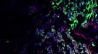
* Fluorescence microscopy: This technique uses fluorescent dyes that bind to specific cellular components, allowing for visualization of those components.
* Electron Microscopy (EM): This type of microscopy uses electrons to illuminate the sample, providing much higher resolution than light microscopy. This allows for the visualization of structures at the nanometer level.
* Transmission electron microscopy (TEM): A beam of electrons is passed through the sample, creating a 2D image of the internal structures.
* Scanning electron microscopy (SEM): A beam of electrons is scanned across the surface of the sample, creating a 3D image of the surface.
2. Staining Techniques:
* Staining involves using dyes to color specific cell components, making them visible under a microscope. Some common stains include:
* Hematoxylin and eosin (H&E) staining: A common histological stain used to visualize the nuclei and cytoplasm of cells.
* Gram staining: Used to differentiate bacteria based on their cell wall structure.
* Immunofluorescence staining: Uses fluorescent antibodies that bind to specific proteins or molecules within the cell.
3. Cell Fractionation:
* This process involves breaking open cells and separating their components based on size and density. This is achieved by:
* Centrifugation: Spins a sample at high speed, separating different components into layers.
* Differential centrifugation: Uses different speeds to isolate specific organelles.
4. Molecular Techniques:
* Immunocytochemistry: Uses antibodies to detect specific proteins within cells.
* In situ hybridization: Uses labeled DNA or RNA probes to detect specific sequences within cells.
* Gene editing techniques: Allow for the manipulation of specific genes within cells, enabling scientists to study the function of these genes.
5. Other Techniques:
* X-ray crystallography: Used to determine the 3D structure of proteins and other molecules.
* Cryo-electron microscopy (cryo-EM): A specialized form of electron microscopy that allows for the visualization of structures at near-atomic resolution.
Choosing the Right Technique:
The choice of technique depends on the specific cell type, the structures to be observed, and the desired level of detail. For example, light microscopy might be sufficient to visualize the overall structure of a cell, while electron microscopy is needed to visualize the detailed structure of organelles.
Note: Each technique has its own limitations and advantages. Researchers often combine multiple techniques to obtain a comprehensive understanding of cell structure and function.