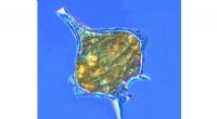
1. Uncovering Ultrastructure:
* High Resolution: Electron microscopes (EM) offer much higher resolution than light microscopes, enabling visualization of structures as small as a few nanometers. This allowed scientists to see internal cell components like ribosomes, mitochondria, Golgi apparatus, and endoplasmic reticulum in exquisite detail, revealing their complex morphology and spatial organization.
* Internal Details: EM allowed the study of organelles in detail, revealing their internal membranes, compartments, and intricate protein machinery. This knowledge was crucial for understanding their specific roles in cellular processes like energy production, protein synthesis, and transport.
* 3D Reconstruction: Techniques like transmission electron microscopy (TEM) and scanning electron microscopy (SEM) allow for the creation of 3D reconstructions of cells, providing a more complete picture of their structure and how different components interact.
2. Understanding Cellular Processes:
* Dynamic Events: EM techniques like freeze-fracture and cryo-electron microscopy (cryo-EM) allowed researchers to study dynamic cellular processes like membrane fusion, protein trafficking, and the formation of cellular junctions. These snapshots of cellular events provided crucial insights into the mechanisms underlying these processes.
* Cellular Interactions: EM allowed scientists to visualize interactions between cells, such as the formation of synapses in the nervous system and cell-cell junctions in tissues. This understanding is critical for comprehending the intricate communication and cooperation between cells.
* Pathology and Disease: EM has been instrumental in understanding the changes in cellular structure and function caused by disease. Studying infected cells, tumor cells, and other diseased cells under EM revealed the molecular basis of various diseases and paved the way for targeted therapies.
3. Advancing Research Tools:
* Immuno-EM: Combining EM with immunogold labeling allows researchers to pinpoint the location of specific proteins within cells, providing crucial information about protein localization and function.
* Cryo-EM: The development of cryo-EM techniques has further revolutionized structural biology, allowing scientists to determine the 3D structures of complex macromolecular assemblies like ribosomes, viruses, and protein complexes with atomic resolution.
In summary:
The electron microscope has been an indispensable tool in cell biology, providing a deeper understanding of the intricate structure and function of cells. Its high resolution, versatility, and constant advancements have enabled scientists to explore cellular processes at unprecedented detail, revealing the incredible complexity and elegance of life at the microscopic level.