Here's a breakdown of how to identify it:
Structure:
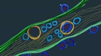
* Network of interconnected membranes: The ER is a continuous system of membranes, forming a vast network within the cell. It's not just a single, isolated structure.
* Sacs and tubules: The ER consists of flattened, sac-like structures called cisternae and interconnected, tube-like structures called tubules.
* Rough ER: Areas of the ER studded with ribosomes are called rough ER and are responsible for protein synthesis and modification.
* Smooth ER: Regions without ribosomes are called smooth ER and are involved in lipid synthesis, detoxification, and calcium storage.
Location:
* Throughout the cytoplasm: The ER extends throughout the cytoplasm, often appearing as a network of interconnected channels.
Key functions:
* Protein synthesis and modification (rough ER)
* Lipid synthesis, detoxification, and calcium storage (smooth ER)
* Transportation of molecules within the cell
* Folding and processing of proteins
Identifying the ER in a cell:
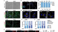
You can identify the ER using electron microscopy, as its network of interconnected membranes is visible. In light microscopy, staining techniques can highlight the ER, making it easier to distinguish from other cellular components.
In summary: The ER is a complex network of membranes that plays a vital role in many cellular processes. Its interconnected structure of sacs and tubules, often studded with ribosomes, distinguishes it within animal cells.