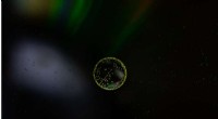
Fluorescence microscopy is a powerful imaging technique that uses fluorescence to visualize specific structures and processes within biological specimens. Here's a breakdown of its key features:
1. Fluorescence: The core principle is based on the phenomenon of fluorescence, where certain molecules (called fluorophores) absorb light at a specific wavelength (excitation wavelength) and then re-emit light at a longer wavelength (emission wavelength).
2. Specific Labeling: Fluorophores are strategically attached to or incorporated into the specimen of interest, allowing researchers to target specific molecules, organelles, or even cellular activities. This selective labeling provides high contrast and specificity, distinguishing the target from its surroundings.
3. Excitation and Emission Filtering: The microscope uses specialized filters to:
* Excitation Filter: Selectively transmits only the excitation wavelength to illuminate the sample.
* Emission Filter: Blocks the excitation wavelength and only allows the emitted fluorescent light to reach the detector.
4. High Sensitivity: The use of fluorescence allows for visualization of extremely small structures and low concentrations of molecules, exceeding the capabilities of conventional light microscopy.
5. Applications: Fluorescence microscopy has revolutionized various fields, including:
* Biology: Studying cellular processes, protein localization, and disease mechanisms.
* Medicine: Diagnosing diseases, monitoring treatment response, and developing new therapies.
* Materials Science: Characterizing materials at the nanoscale level.
In summary, fluorescence microscopy is a versatile and powerful technique that allows scientists to visualize specific targets within a complex biological system by exploiting the unique properties of fluorescence. It offers high sensitivity, specificity, and versatility, making it an indispensable tool for research and diagnostics.