1. Light Microscope (LM)
* Appearance: You'll see the basic shape and structure of the algae cell.
* Chloroplasts: Appear as green, oval or disc-shaped structures within the cell.
* Cell Wall: The cell wall might be visible as a thin, outlining line.
* Nucleus: May be visible as a darker spot within the cytoplasm.
* Other Structures: Vacuoles (fluid-filled sacs) and other organelles may be vaguely discernible.
* Magnification: Typically up to 1000x.
* Resolution: Limited. You won't be able to see fine details of internal structures.
2. Compound Microscope (LM)
* Appearance: Similar to a standard light microscope, but with improved resolution.
* Chloroplasts: More clearly defined.
* Cell Wall: More visible.
* Internal structures: May be seen with more clarity.
* Magnification: Can reach 1500x or higher.
* Resolution: Better than a basic light microscope.
3. Phase-Contrast Microscope (LM)
* Appearance: Provides greater contrast, making internal structures easier to distinguish.
* Chloroplasts: Appear very distinct with a high degree of contrast.
* Cell Wall: More apparent.
* Internal structures: Appear with more clarity.
* Magnification: Similar to other light microscopes.
* Resolution: Better than a standard light microscope, especially for observing internal structures.
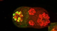
4. Fluorescence Microscope (FM)
* Appearance: Uses fluorescent dyes to highlight specific structures within the algae cell.
* Chloroplasts: Can be made to fluoresce in various colors, depending on the dye used.
* Cell Wall: May be stained with fluorescent dyes.
* Other structures: Specific organelles can be identified based on their fluorescence.
* Magnification: Similar to other light microscopes.
* Resolution: Similar to other light microscopes, but with enhanced visualization of specific structures.
5. Scanning Electron Microscope (SEM)
* Appearance: Provides a 3D, surface view of the algae cell.
* Cell Wall: Detailed surface textures and patterns are visible.
* Surface Structures: Any projections or appendages on the cell's surface are clearly shown.
* Magnification: Can reach 100,000x or more.
* Resolution: Very high, allowing for detailed surface structure analysis.
6. Transmission Electron Microscope (TEM)
* Appearance: Provides a cross-section view of the algae cell, showing internal structures in great detail.
* Chloroplasts: Internal membranes and grana are clearly visible.
* Cell Wall: The layers and composition of the cell wall can be observed.
* Internal Structures: Organelles, such as the nucleus, ribosomes, and endoplasmic reticulum, are visualized in fine detail.
* Magnification: Can reach millions of times.
* Resolution: Extremely high, allowing for the visualization of individual molecules and their arrangement within the cell.
In summary:
* Light Microscopes: Provide basic shapes, but limited internal detail.
* Phase-Contrast Microscopes: Enhance internal structure visibility.
* Fluorescence Microscopes: Highlight specific structures using fluorescent dyes.
* Electron Microscopes (SEM and TEM): Offer incredibly high magnification and resolution, revealing surface detail (SEM) or internal ultrastructure (TEM).
The choice of microscope depends on the specific question you're trying to answer about the algae cell.