Here's a breakdown:
Microscopy is a broad term referring to the use of a microscope to view small objects or structures that are not visible to the naked eye.
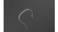
Live cell imaging or live-cell microscopy refers to the techniques used to observe and study living cells in real-time under a microscope. This can involve:
* Bright-field microscopy: This is the most basic type of microscopy, where light is shone through the sample and the image is observed in transmitted light.
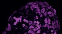
* Fluorescence microscopy: This technique uses fluorescent dyes or proteins to label specific cellular components, allowing for the visualization of specific structures within the cell.
* Phase-contrast microscopy: This technique enhances the contrast of transparent objects by using a phase plate to shift the phase of light passing through the sample.
* Differential interference contrast (DIC) microscopy: Similar to phase-contrast microscopy, DIC microscopy uses polarized light to enhance the contrast of transparent objects.
* Confocal microscopy: This technique uses a laser to scan the sample point-by-point, creating a 3D image of the sample.
Why study living cells under a microscope?
Live-cell imaging allows scientists to:
* Observe cellular processes in real-time: This includes processes like cell division, migration, and protein synthesis.
* Study the dynamics of cellular structures: This can reveal how structures move, change shape, and interact with other cellular components.
* Investigate the effects of drugs and other treatments on cells: This is important for understanding how drugs work and developing new treatments.
* Identify and track specific cells: This is useful for studying the development of tissues and organs.
Live-cell imaging is a powerful tool for studying the fundamental processes of life and for understanding the workings of cells in health and disease.