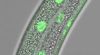
1. Enhancing Contrast and Visibility:
* Most cells are nearly transparent, making it difficult to distinguish their structures under a microscope.
* Staining adds color and contrast to different cellular components, making them more visible.
* Different stains target specific structures like the nucleus, cytoplasm, or cell membrane, allowing us to visualize their unique features.
2. Identifying Specific Cellular Components:
* Different stains have an affinity for specific molecules or structures within cells.
* For example, hematoxylin stains the nucleus blue, while eosin stains the cytoplasm pink.
* This allows researchers to identify and differentiate various cellular components.
3. Analyzing Cell Morphology and Function:
* Staining patterns can reveal information about cell morphology, including size, shape, and the presence of organelles.
* Certain staining techniques can also highlight specific cellular functions, such as the presence of enzymes or proteins.
4. Diagnosing Diseases:
* In medical settings, staining is crucial for diagnosing diseases.
* For instance, Pap smears use stains to detect abnormal cells that may indicate cervical cancer.
* Biopsies often involve staining to identify the presence of cancerous cells.
5. Research and Development:
* Staining plays a vital role in research, allowing scientists to study cell structure, function, and interactions.
* It helps researchers understand cellular processes and develop new treatments for diseases.
In summary, staining cells under a microscope is essential for:
* Improving visibility and contrast.
* Identifying specific cellular components.
* Analyzing cell morphology and function.
* Diagnosing diseases.
* Supporting research and development.