1. Enhanced Visibility:
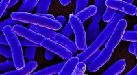
* Contrast: Most microbial cells are transparent and difficult to see under a light microscope. Stains provide contrast, making them stand out against the background. This makes it easier to observe their shape, size, and arrangement.
* Detail: Some stains can highlight specific structures within the cell, such as the cell wall, capsule, flagella, or internal organelles. This provides more detailed information about the cell's composition and function.
2. Differentiation:
* Gram Staining: This is a widely used differential stain that distinguishes between two major types of bacteria: gram-positive and gram-negative. This differentiation is crucial for understanding bacterial cell wall structure and selecting appropriate antibiotics.
* Acid-fast Staining: This technique differentiates bacteria based on their ability to retain a dye even after treatment with acid. It is used to identify species like Mycobacterium tuberculosis, which have a waxy cell wall.
3. Identification and Diagnosis:
* Specific Stains: Certain stains are designed to target specific components of microbes, allowing for their identification. For example, endospore stains can help in detecting endospores, which are highly resistant structures formed by some bacteria.
* Clinical Diagnosis: Stains are widely used in clinical microbiology for diagnosing infections. For example, a Gram stain of a sputum sample can help in identifying the causative bacteria of pneumonia.
4. Studying Cell Processes:
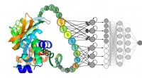
* Fluorescence Microscopy: Fluorescent dyes can be used to label specific molecules within the cell, allowing researchers to track and study cellular processes in real-time. This is particularly useful for studying cell division, protein trafficking, and other dynamic events.
In summary, stains are essential tools for microbiologists as they provide enhanced visibility, allow for differentiation between different types of microbes, facilitate identification and diagnosis, and aid in studying various cellular processes.