Here's how it works:
1. Primary Stain: The first dye is applied to the sample. This dye will bind to specific structures based on their chemical properties.
2. Decolorization: A decolorizing agent is used to remove the primary stain from certain structures, while leaving it in others.
3. Counterstain: A second dye with a contrasting color is applied. This dye will bind to structures that were decolorized in the previous step.
The result of this process is a sample where different cell types or structures appear in different colors. This allows researchers to easily differentiate between them and study their characteristics.
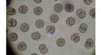
Examples of differential stains:
* Gram stain: Used to differentiate between bacteria based on their cell wall structure (Gram-positive vs Gram-negative).
* Acid-fast stain: Used to identify bacteria that have a waxy cell wall, such as Mycobacterium tuberculosis.
* Ziehl-Neelsen stain: Similar to the acid-fast stain, used to identify Mycobacterium species.
* Giemsa stain: Used to stain blood cells, identifying different types of white blood cells.
* Wright's stain: Similar to Giemsa, used for blood cell staining.
Advantages of differential stains:
* Improved visualization: Different colors make it easier to identify and study specific structures.
* Classification: Can be used to classify bacteria, blood cells, and other biological specimens.
* Diagnosis: Can be used to diagnose diseases based on the presence or absence of specific microorganisms.
Overall, differential staining is a powerful tool used in microbiology, hematology, and other fields to improve the visualization, classification, and diagnosis of biological samples.