1. Light Microscopes:
* Bright-field microscope: The most common type, uses visible light to illuminate the sample.
* Dark-field microscope: Illuminates the sample from the sides, creating a bright image against a dark background, good for observing unstained specimens.
* Phase-contrast microscope: Enhances the contrast of transparent specimens by manipulating the phase of light passing through them.
* Differential interference contrast (DIC) microscope: Similar to phase-contrast but uses polarized light to create a 3D-like image.
2. Electron Microscopes:
* Transmission electron microscope (TEM): Uses a beam of electrons to create an image of the internal structure of thin specimens.
* Scanning electron microscope (SEM): Scans a focused beam of electrons across the surface of a sample to create detailed images of the topography.
3. Other Microscopes:
* Fluorescence microscope: Uses fluorescent dyes to illuminate specific structures or molecules within a sample.
* Confocal microscope: A type of fluorescence microscope that uses a pinhole to eliminate out-of-focus light, creating sharper images of thick specimens.
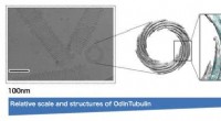
* Atomic force microscope (AFM): Uses a sharp probe to scan the surface of a sample, creating a 3D image of the surface at the atomic level.
* Scanning probe microscope (SPM): A general term for a group of microscopes that use a probe to scan a sample's surface, including AFM, STM (scanning tunneling microscope), etc.
Note: This is not an exhaustive list, and there are many more specialized types of microscopes used in research and industry.