Based on Illumination Source:
* Light Microscopes:
* Bright-field microscope: Most common type, uses visible light to illuminate the sample.
* Dark-field microscope: Illuminates the sample from the sides, making the specimen appear bright against a dark background.
* Phase-contrast microscope: Uses differences in refractive index to enhance contrast in transparent specimens.
* Differential interference contrast (DIC) microscope: Similar to phase-contrast but provides a more 3D-like image.
* Polarized light microscope: Uses polarized light to study materials that exhibit birefringence (different refractive indices in different directions).
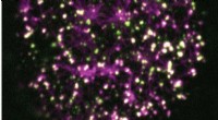
* Fluorescence microscope: Uses fluorescent dyes to illuminate specific structures within the sample.
* Confocal microscope: Uses lasers and a pinhole to create sharp images of thick specimens.
* Electron Microscopes:
* Transmission electron microscope (TEM): Uses a beam of electrons to create images of extremely thin samples, providing high resolution.
* Scanning electron microscope (SEM): Uses a focused beam of electrons to scan the surface of a sample, creating detailed 3D images.
* Scanning transmission electron microscope (STEM): Combines the principles of TEM and SEM.
Other Types:
* Scanning probe microscope (SPM): A family of microscopes that uses a sharp tip to scan the surface of a sample and create a detailed image.
* Atomic force microscope (AFM): One of the most common SPMs, can image individual atoms.
* Scanning tunneling microscope (STM): Another common SPM, used to study the surface of conductive materials.
* Acoustic microscope: Uses sound waves to create images of materials.
* X-ray microscope: Uses X-rays to create images of thick samples, providing high penetration power.
Specialized Microscopes:
* Super-resolution microscopy: A family of techniques that exceed the diffraction limit of light microscopes, allowing visualization of structures smaller than 200 nm.
* Light sheet microscopy: A technique that illuminates a thin sheet of the sample, reducing photobleaching and allowing for 3D imaging.
* Holographic microscopy: Uses holographic techniques to create 3D images of samples.
Note: This is not an exhaustive list, and there are many other specialized microscopes developed for specific applications.
It's important to remember that the choice of microscope depends on the specific application and the type of sample being examined.