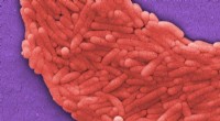
Flagellar staining is a technique used to visualize bacterial flagella, which are thin, whip-like appendages that help bacteria move. The principle behind this staining is based on increasing the diameter of the flagella so that they become visible under a light microscope.
Here's how it works:
1. Mordant: A mordant is a chemical substance that binds to the flagella and creates a larger surface area. This can be done by using a solution like tannic acid or potassium alum.
2. Staining: After the mordant, a stain like silver nitrate or pararosaniline is applied. This stain will adhere to the mordant and create a thick layer around the flagella, making them much more visible.
3. Washing: The excess stain is then washed away, leaving only the stained flagella visible against the background.
Key points about flagellar staining:
* Sensitivity: This technique is very sensitive, as it can detect the presence of flagella even if they are present in small numbers.
* Specificity: Flagellar staining is highly specific for flagella. It does not stain other bacterial structures.
* Types: There are several different methods of flagellar staining, each with its own specific protocol and reagents.
* Applications: Flagellar staining is used in microbiology for:
* Identifying bacterial species: The number and arrangement of flagella can be used to help identify different types of bacteria.
* Studying bacterial motility: Flagellar staining helps to observe how bacteria move and interact with their environment.
* Research: This technique is also used in research to study the structure and function of bacterial flagella.
Overall, flagellar staining is a powerful tool that allows us to visualize these important bacterial structures and learn more about their role in bacterial biology.