Microscopes are essential tools for visualizing the microscopic world, ranging from cells to intricate structures within them. Here's a breakdown of common microscope types and their functions:
1. Light Microscopes (Optical Microscopes):
* Bright-Field Microscopy: The most basic type, where light passes through the specimen and is projected onto the eye or a camera. It's good for viewing stained specimens and general observation.
* Dark-Field Microscopy: Uses a special condenser to only allow light scattered by the specimen to reach the objective. Creates a bright image against a dark background, ideal for viewing unstained, transparent specimens.
* Phase-Contrast Microscopy: Exploits the differences in refractive index between different parts of a specimen. Enhances contrast in unstained specimens, revealing details like cell organelles.
* Differential Interference Contrast (DIC) Microscopy: Similar to phase-contrast, but uses polarized light to create a 3D-like effect. Provides excellent contrast and detail in unstained specimens.
* Fluorescence Microscopy: Uses fluorescent dyes that absorb light at one wavelength and emit it at another. Ideal for visualizing specific molecules or structures within cells.
2. Electron Microscopes:
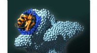
* Transmission Electron Microscopy (TEM): Uses a beam of electrons to illuminate a very thin specimen. Offers high resolution and the ability to view internal structures of cells.
* Scanning Electron Microscopy (SEM): Scans the surface of a specimen with a focused electron beam. Provides detailed images of the specimen's topography and surface structures.
3. Other Specialized Microscopes:
* Confocal Microscopy: Uses lasers to scan a specimen, creating 3D images by eliminating out-of-focus light. Good for viewing thick specimens and for studying dynamic processes.
* Super-Resolution Microscopy: A set of techniques that overcome the diffraction limit of light, allowing for resolutions beyond the capabilities of traditional light microscopy.
* Atomic Force Microscopy (AFM): Uses a sharp probe to scan the surface of a specimen, providing extremely high resolution images. Can be used to study a variety of materials, from polymers to biological samples.
4. Microscope Applications:
* Biology & Medicine: Studying cells, tissues, and microorganisms, diagnosing diseases, research.
* Materials Science: Analyzing material properties, identifying defects, developing new materials.
* Engineering & Manufacturing: Quality control, inspecting tiny components, characterizing surfaces.
* Forensic Science: Analyzing evidence, identifying substances, reconstructing events.
Choosing the right microscope depends on the specific application and the sample being studied. Each type has its own advantages and disadvantages, so it's essential to consider the desired resolution, magnification, and the characteristics of the specimen.