1. Electrophoresis System:
* Gel: The most common types are agarose and polyacrylamide. Agarose gels are used for larger molecules, while polyacrylamide gels are used for smaller molecules.
* Buffer: An electrolyte solution that conducts electricity and maintains the pH of the gel.
* Power Supply: Provides the electrical current that drives the migration of molecules.
* Comb: Creates wells in the gel where samples are loaded.
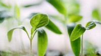
2. Sample Preparation:
* Sample: This can be DNA, RNA, proteins, or other molecules that you want to separate.
* Loading Dye: Contains a tracking dye that allows you to monitor the progress of electrophoresis and a dense solution to help the sample sink into the well.
3. Detection and Analysis:
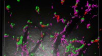
* Stain: Used to visualize the separated molecules. Common stains include:
* Ethidium Bromide (EtBr) for DNA and RNA (fluoresces under UV light)
* Coomassie Blue for proteins
* Imaging System: A system that can capture and analyze the stained gel, such as a UV transilluminator, gel documentation system, or scanner.
* Software: For analyzing the results, measuring band sizes, and calculating molecular weights.
4. Other Equipment:
* Pipettes: Used to accurately load samples into the gel wells.
* Micropipette: A small pipette that measures very small volumes of liquid.
* Gloves: To protect your hands from chemicals and stains.
Key Measurements in Electrophoresis:
* Migration Distance: The distance traveled by a molecule from the well to its final position on the gel.
* Mobility: A measure of how fast a molecule moves in an electric field.
* Molecular Weight: The size of the molecule, which can be estimated based on its migration distance relative to known standards.
* Concentration: The amount of a molecule present in a sample, which can be measured by the intensity of the stained band.
By using this equipment and following established protocols, scientists can effectively separate and analyze biomolecules using electrophoresis.