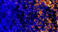
Enhanced Contrast:
- Dark-field microscopy provides superior contrast compared to bright-field microscopy, allowing better visualization of unstained or transparent specimens.
Illumination Technique:
- In dark-field microscopy, the light source is positioned at an oblique angle to the specimen, creating a cone of illumination. This illumination technique prevents direct light from reaching the objective lens, resulting in a dark background.
Scattering Effect:
- The light from the oblique illumination scatters when it interacts with minute structures or particles in the specimen. This scattering effect generates a bright halo or ring around the edges of the objects, making them stand out against the dark background.
Detection of Ultra-small Structures:
- Dark-field microscopy excels in detecting extremely small structures or particles that are difficult to visualize using bright-field microscopy. It enables the observation of tiny details and features, such as bacterial flagella, viruses, and subcellular structures.
Examination of Unstained Specimens:
- Dark-field microscopy allows for the examination of unstained specimens, which is particularly useful for observing living cells or microorganisms without the need for potentially harmful staining procedures.
Enhanced Visualization of Transparent Objects:
- Transparent objects or structures, which may appear almost invisible under bright-field microscopy, become more visible due to the scattering effect in dark-field microscopy.
Complementary Technique:
- Dark-field microscopy complements other microscopy techniques by providing different information and insights about the specimen. It can be used in conjunction with bright-field microscopy to obtain a comprehensive understanding of the sample.
Real-Time Observation:
- Dark-field microscopy enables real-time observation of dynamic processes and movements within living cells or microorganisms, as it does not require specimen fixation or staining.
However, it's worth noting that dark-field microscopy also has limitations, such as reduced image resolution compared to bright-field microscopy and potential challenges in distinguishing between different structures based solely on their scattering patterns. Therefore, the choice of microscopy technique depends on the specific requirements and objectives of the research or observation being conducted.