1. Chromosome Shape and Orientation:
- Chromosomes have a complex three-dimensional structure that can vary depending on the species, cell type, and stage of cell division.
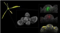
- In lateral view, chromosomes are visualized along their longitudinal axis, showing their full length and allowing for the observation of chromosome arms, centromeres, and telomeres.
- In polar view, chromosomes are seen from above, providing a cross-sectional view that captures the overall shape and arrangement of the chromosomes within the metaphase plate.
2. Centromere Positions:
- The centromere is the region of the chromosome where spindle fibers attach during cell division.
- In lateral view, the centromere is typically visible as a constriction or narrowing point along the chromosome's length.
- In polar view, the centromeres of different chromosomes appear as distinct dots or foci when viewed from above the metaphase plate.
3. Chromatid Overlapping:
- During metaphase, sister chromatids, which are exact copies of each other, are held together by the cohesion complex.
- In lateral view, the chromatids may be somewhat separated, allowing for the visualization of individual chromatid arms.
- In polar view, the sister chromatids often overlap more extensively, obscuring some details of their individual structures.
4. Depth of Field and Resolution:
- Microscopy techniques have limitations in terms of depth of field, which affects the range of focus within an image.
- In lateral view, the focal plane can be adjusted to capture different regions of the chromosome along its length, providing more detailed information about specific areas.
- In polar view, the focal plane is typically set to capture the entire metaphase plate, resulting in a composite image that includes chromosomes at different depths. This can contribute to differences in the level of detail observed between the two views.
In summary, the differences between lateral and polar views of chromosomes during metaphase arise from variations in chromosome orientation, centromere positions, chromatid overlapping, and limitations in microscopy techniques. Each view provides complementary information about chromosome structure and organization, allowing researchers to gain a comprehensive understanding of chromosome behavior during cell division.