Iron is essential for various physiological functions, including oxygen transport, energy production, and DNA synthesis. Cells have developed specialized mechanisms to ensure an adequate supply of iron from the external environment. The process of iron absorption occurs primarily in the small intestine, where specialized epithelial cells play a crucial role.
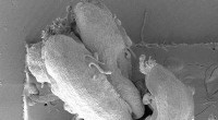
The electron microscope images provide a detailed look at the cellular structures and molecules involved in iron absorption. Here are some key features observed:
1. Microvilli: The intestinal epithelial cells possess numerous microvilli, which are finger-like projections that increase the surface area for nutrient absorption. These microvilli are densely packed and covered in glycocalyx, a layer of carbohydrates that helps capture iron ions.
2. Iron Transporters: The apical membranes of intestinal epithelial cells express specific iron transporters, such as divalent metal transporter 1 (DMT1) and ferroportin. DMT1 facilitates the uptake of ferrous iron (Fe2+) from the intestinal lumen into the enterocytes, while ferroportin is responsible for iron export from the cells into the bloodstream.
3. Intracellular Vesicles: Once inside the enterocytes, iron is stored temporarily in intracellular vesicles called endosomes. These vesicles are specialized compartments that regulate the trafficking and distribution of iron within the cells.
4. Ferritin: Iron is also stored in the form of ferritin, a protein complex that sequesters iron in a non-toxic and readily releasable form. Ferritin is found in both the cytoplasm and mitochondria of intestinal epithelial cells.
5. Basolateral Export: Iron that is not immediately required by the enterocytes is exported across the basolateral membrane into the bloodstream. This process is facilitated by ferroportin, which transports iron out of the cells in response to systemic iron demand.
The electron microscope images not only reveal the cellular structures involved in iron absorption but also provide insights into the molecular mechanisms that regulate this process. By understanding the intricate details of iron uptake, researchers can gain a deeper understanding of iron homeostasis, iron-related disorders, and develop potential therapeutic strategies to address iron deficiency and related conditions.