ATP serves as the primary energy currency for cells, providing the necessary fuel to power various biological reactions. It consists of a molecule of the sugar ribose attached to three phosphate groups. When cells require energy, they break down ATP, liberating the energy stored within the phosphate bonds and releasing ADP (adenosine diphosphate) as a byproduct.
However, the precise mechanisms by which cells achieve this crucial energy-yielding process have remained elusive, hindering our full understanding of cellular function. In this landmark study, the MIT research team employed a combination of cutting-edge microscopy techniques and computational modeling to capture and analyze the events occurring at the molecular level during ATP breakdown.
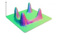
Using a custom-built microscope, the researchers were able to visualize the intricate interactions between ATP molecules and a key enzyme responsible for cleaving the phosphate bonds, known as ATP synthase. Their real-time imaging revealed the precise choreography of molecular movements that occur during the breakdown process.
Additionally, computational modeling allowed the researchers to simulate and analyze the behavior of ATP molecules within cells. By integrating the experimental observations with computational data, they could develop a comprehensive understanding of the underlying physical principles governing the breakdown of ATP.
The findings of this study have significant implications for our knowledge of cellular energy metabolism and may inform future research into various human diseases and disorders associated with energy production. By unraveling the intricate details of this fundamental process, the work contributes to our broader understanding of life's intricate mechanisms and could pave the way for the development of novel therapeutic strategies targeting energy metabolism.