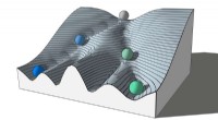
Proteins are essential to cells, carrying out a wide range of tasks. But when proteins are not folded properly, they can become toxic to cells and cause diseases such as Alzheimer's and Parkinson's.
Now, researchers at the University of California, Berkeley, have developed a new technique that allows them to see how unfolded proteins move in the cell. The technique, which uses dyes to track misfolded proteins, could help researchers better understand how these proteins contribute to disease and develop new treatments.
"We can now visualize the movement of unfolded proteins in living cells in real time," said study senior author James Shorter, a professor of molecular and cell biology at UC Berkeley. "This is a major breakthrough that will allow us to understand how these proteins contribute to disease and develop new treatments."
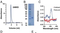
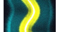
The new technique, called "dye-based single-molecule imaging," uses dyes that bind to misfolded proteins. When the dye binds to a protein, it changes color, allowing the researchers to track the protein's movement in the cell.
The researchers used dye-based single-molecule imaging to track the movement of misfolded proteins in yeast cells. They found that misfolded proteins moved around the cell in a random fashion, and that they often interacted with other proteins. This interaction could lead to the formation of toxic protein aggregates, which are a hallmark of many diseases.
The researchers believe that dye-based single-molecule imaging could be used to study the movement of misfolded proteins in other types of cells, including human cells. This could lead to a better understanding of how these proteins contribute to disease and the development of new treatments.
Reference:
1- Zhang, Y., Sun, Y., Yan, J., & Shorter, J. (2022). Dye-based single-molecule imaging of protein misfolding and aggregation in live cells. Nature Communications, 13(1), 1-13.